Anatomy Next: The 3D human atlas
This article was first published in late 2017 in CoFounder No 10.
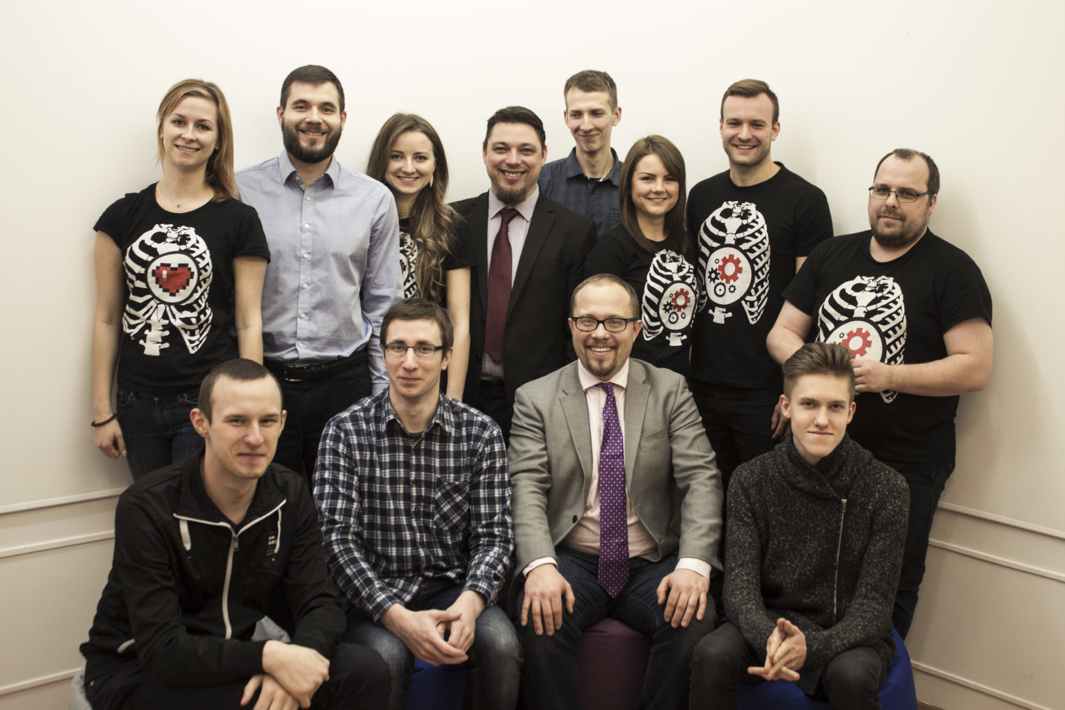
Anatomy Next changes anatomy classes for medical students. The team has completed a digital atlas of the human head and neck, along with detailed 2D maps and interactive 3D visuals of the cranial nerves. They plan to have the whole male body completed by spring 2018.
The team expects their 3D simulations to turn the mindless memorisation of textbooks (which is forgotten soon after an exam) into a real understanding of human anatomy that students will carry into their professional lives.
The company was started by a Latvian sculptor, Uldis Zarins, who repeatedly failed to correctly sculpt the cheek of a classical sculpture. He tried every trick in the book but could not find a way to measure the vast round area. Teachers sent him to the library, where Uldis tried every book and eventually started deconstructing each element of the human body into basic shapes and posting his discoveries on Facebook.
A successful Kickstarter campaign, together with his co-founder, Sandis Kondrats, helped them publish the first modern anatomy book for sculptors, available at anatomy4sculptors.com. When the two discovered that the digital generation of medical students was still learning anatomy from books written in the 18th century, they started to ponder the opportunities in digital and Anatomy Next was born.
The timing couldn’t have been better. Today, teachers who spend one hour talking about a complicated concept, while reading from their slides, are competing with bright, charismatic YouTubers who complete the same explanation – with witty visuals – in three minutes. The pressure to remain relevant makes universities more open to considering solutions like Anatomy Next.

Based on high-resolution radiological data, the 3D Anatomy Atlas allows students to experience anatomical structures in the finest detail. Students can fully manipulate each structure, using web-based, standalone or web application platforms to rotate, zoom in and view isolated muscles, bones, nerves and blood vessels. The application offers options to hide and fade structures, to see their location in relation to other tissues, bones and important landmarks. Detailed educational descriptions allow students to discover muscle origin, insertion, function, innervation and blood supply. Anatomy Next currently has pilot programmes at seven universities in the US and EU, with 50 more in negotiation.
The team continues to integrate the newest technology for enhanced experiential learning. They use augmented reality and virtual reality, aiming to produce a learning experience as close to real life as possible. If successful, this could mean that surgeons would graduate from the Anatomy Next program already knowing what it feels like to put together any combination of broken bones, severed nerves or torn muscles. The team expects that this can help decrease the share of fatalities and disabilities due to medical errors. They already have over 7,000 downloads to their HoloLens application and are working with Microsoft to make VR more relevant in medicine.


